The prostate MRI
Prostate cancer is common: around one in five men will develop prostate cancer in the course of their life. While prostate palpation by the doctor’s finger is still the only examination paid for by German health insurance companies – although there is not a single study showing that DRU (digital rectal examination) has ever saved a single life – modern diagnostics have evolved.
Prostate MRI (magnetic resonance imaging – MRI) detects prostate cancer with over 90% certainty – or excludes relevant cancer foci. Many experts are now of the opinion that an MRI of the prostate should be carried out first if a prostate carcinoma is suspected.
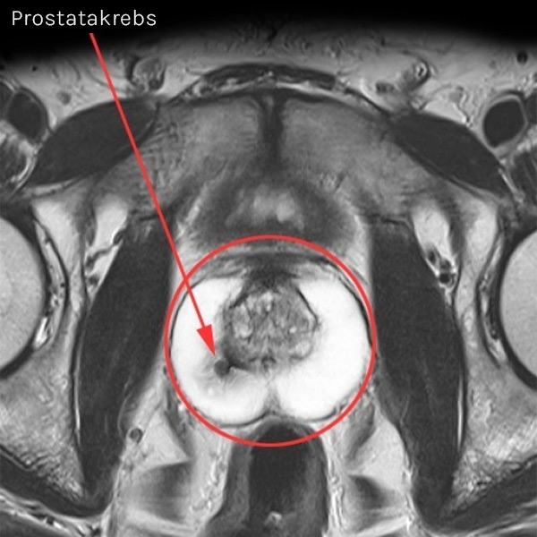
Small prostate cancer focus (arrow) in the right outer zone of the prostate. If detected early, the cancer can be treated easily and without side effects with focal therapy, e.g. using irreversible electroporation (IRE – NanoKnife). This small focus would not have been noticed during a palpation examination, as it is embedded in normal prostate tissue (light color in the image).
If no suspicious lesions are found in the MRI, a biopsy, the invasive removal of tissue samples from the prostate, is often unecessary. If suspicious foci are present, the biopsy can be precisely targeted to the tumor using MRI – either by fusion biopsy or, even more precisely, directly in the MRI scanner under image control.

MRI of the prostate is, amongst others, recommended by:
- European Association of Urology (EAU)
- American Urological Association (AUA)
- National Comprehensive Cancer Network (NCCN)
Even for the early detection of prostate cancer, the prostate MRI has proven to be by far the best method. It is far superior to palpation (DRU), the PSA test and ultrasound examinations.
Our brochure summarizes the key points on prostate cancer screening and diagnosis in an easy-to-understand way. Weitere Informationen zu diesem Thema finden sie unter vitusprivatklinik.com. Our experts would also be happy to advise you.


