Thyroid nodules are relatively common; some of the nodules may conceal cancerous lesions. Differentiating between benign and malignant lumps has been difficult up to now: neither nuclear medicine scintigraphy nor sonography (ultrasound) can reliably rule out cancerous lesions. Even tissue biopsies often yield inconclusive results. With diffusion-weighted MRI, it is now possible to differentiate between benign and malignant thyroid nodules non-invasively.
Special, so-called diffusion-weighted sequences have been used for several years to distinguish malignant from benign changes in the body. This is possible because in malignant tumors – cancer – the mobility of water molecules is restricted by the smaller extracellular space – a specific difference to benign changes that can be visualized by diffusion-weighted MRI sequences. The so-called multiparametric MRI (mp-MRI) examination, combined with MR sequences that measure contrast uptake and wash-out in tissue, along with high-resolution MR images depicting structural details, demonstrates high sensitivity and specificity for detecting or ruling out malignant tumors, not only in the thyroid gland.
These multiparametric MRI examinations are also employed in tumor diagnostics for head and neck tumors, as well as for detecting changes in the lungs, liver, pancreas, female breast, and male prostate.
Risk factors
- Iodine deficiency
- Family history
- Autoimmune diseases
- Radiation exposure
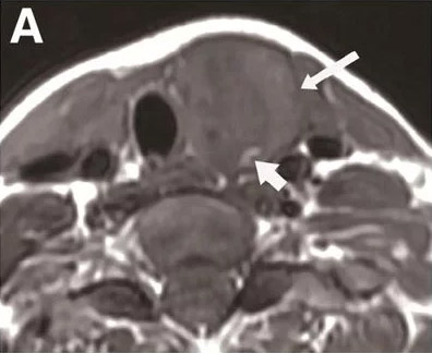
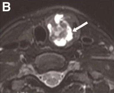
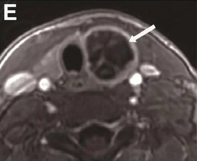
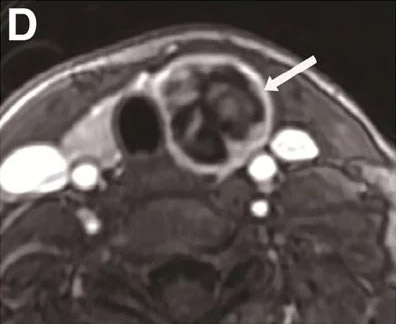
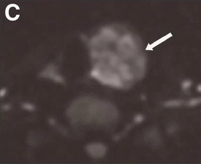
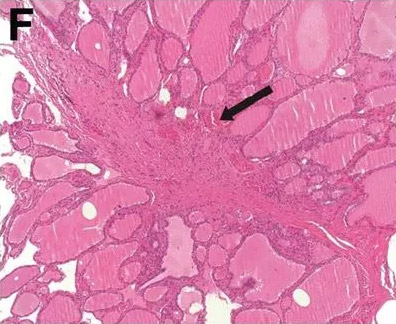
MRI images of a multiparametric examination (A – E) in a 47-year-old woman with a typical heterogeneous thyroid nodule; F shows the histopathology.
In heterogeneous nodules such as these, it is almost impossible to detect a cancer focus through biopsy. It is therefore almost impossible to rule out cancer.
In this case, despite the different tissue components in the thyroid nodule, the multiparametric MRI provided no evidence of carcinoma – which was confirmed by the histological examination of the nodule.


