The paranasal sinuses are a site of frequent inflammation. While X-rays used to be taken to visualize changes in the nasal sinuses, this has not been recommended for years.
Computed tomography (CT) or magnetic resonance imaging (MRI) provide 3D cross-sectional images with superior contrast and detail resolution. A CT scan is recommended if an operation is pending and the fine bone structures of the paranasal sinuses need to be visualized in detail.
In all other cases, MRI is superior because it shows inflammation and tumors better than CT and does not expose the patient to radiation.
Important: Sinusitis is not to be trifled with! Chronic inflammation of the paranasal sinuses can lead to a progression of the infection. The chronic inflammation descends from the level of the paranasal sinuses down into the lungs, leading to chronic bronchitis, which can result in severe and irreversible changes in the lungs that significantly impair lung function.
Our recommendation
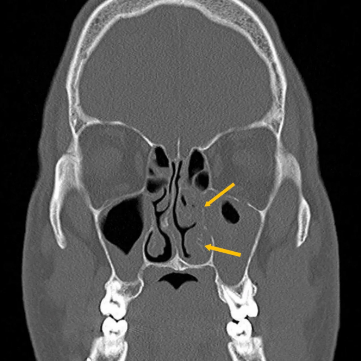
A CT scan of the paranasal sinuses provides an accurate diagnosis of sinusitis and other inflammations or structural problems.
A special type of computer tomography (CT)
Digital volume tomography (DVT) versus CT
A special form of CT examination of the paranasal sinuses is DVT, digital volume tomography. However, it can only image the bone structures with sufficient accuracy, while CT can also image the soft tissue. It is often claimed that DVT has the advantage of lower radiation exposure. This is not physically correct. To achieve the same image quality, the same beam energy must be applied per unit volume. However, this is lower in the high-contrast range, i.e. when imaging the bones, than when the soft tissues are also to be imaged. If the CT is only intended to image the bones, as is the case with CBCT, the radiation exposure of both procedures is the same, but the CT is more precise (see image).

CT image of the paranasal sinuses in chronic sinusitis with incipient destruction of the bones and turbinates (arrows).


